We analyze your cell and tissue samples using light or immunofluorescence/laser scanning microscopy.
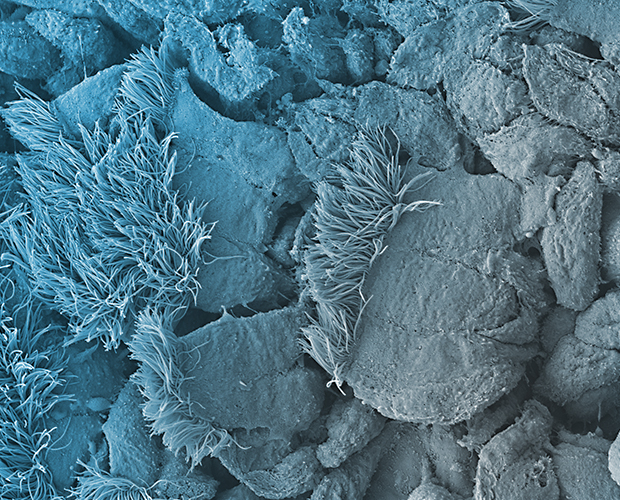
Using high-speed microscopy, we analyze, for example, the ciliary beating of viable tissue models of the conductive airways.
Using high-speed microscopy, we analyze, for example, the ciliary beating of viable tissue models of the conductive airways.

We analyze your cell and tissue preparations using light or immunofluorescence/laser scanning microscopy. Using the light microscope, we can extract defined areas from tissue sections via laser microdissection in order to analyze them in the next step, e.g. using single cell PCR. On the ultrastructural level, we offer analyses at the transmission (with Cryo Ion Slicer) or scanning electron microscope with optional elemental analysis. Furthermore, we perform cross-section preparations with ion beam techniques (Focused Ion Beam, Cryo-Cross-Section Polishing).

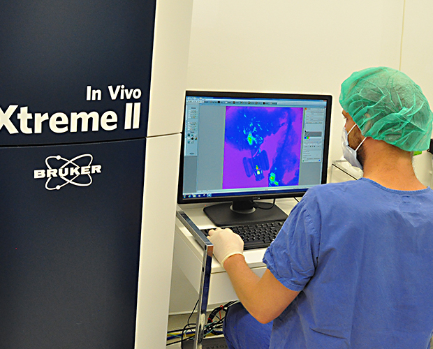
Using high-speed microscopy, we analyze, for example, the cilia beat of viable tissue models of the conducting airways. An in vivo imaging system (IVIS) can be used to non-invasively study 3D tissues or live small animals (e.g., mice, rats, rabbits). Animals can be held in the instrument using inhalation anesthesia for short periods of time during the analyses. Fluorescence, luminescence as well as X-ray images can be performed and quantified. Application examples can be found e.g. in the field of tumorigenicity or implantation studies.